
What is Endoscopic Ultrasound (EUS)?
Endoscopic Ultrasound (EUS) is a advanced diagnostic procedure that combines endoscopy and ultrasound technology. It uses a thin, flexible tube called an endoscope equipped with a small ultrasound probe at its tip. This allows doctors to obtain high-resolution images of the inner lining of the digestive tract as well as the surrounding organs and tissues that are not easily visible through standard tests.
Unlike traditional ultrasound performed from outside the body, EUS brings the imaging device very close to the target area inside the gastrointestinal (GI) tract. This proximity produces clearer, more detailed pictures of structures such as the esophagus, stomach, duodenum, colon, rectum, pancreas, liver, gallbladder, and nearby lymph nodes.
Uses and Benefits of EUS
EUS plays a crucial role in evaluating various digestive health issues. It is particularly valuable for:
Diagnosing and staging cancers
Including cancers of the esophagus, stomach, pancreas, rectum, and even lung cancer by assessing tumor size, depth of invasion, and spread to lymph nodes.
Evaluating pancreatic conditions
Such as cysts, tumors, chronic pancreatitis, or acute inflammation.
Detecting bile duct and gallbladder problems
Including stones, strictures, or blockages.
Assessing subepithelial lesions
Growths beneath the lining of the GI tract
Investigating unexplained symptoms
Like abdominal pain, weight loss, or abnormal findings on other imaging tests (CT or MRI).
The key benefits include high accuracy due to detailed imaging, the ability to perform biopsies in the same session, minimal discomfort, and quicker recovery compared to surgical alternatives. It supports early detection and precise treatment planning, especially in complex gastrointestinal and oncosurgical cases.
How Does EUS Work?
There are two main approaches to gallbladder removal:
1
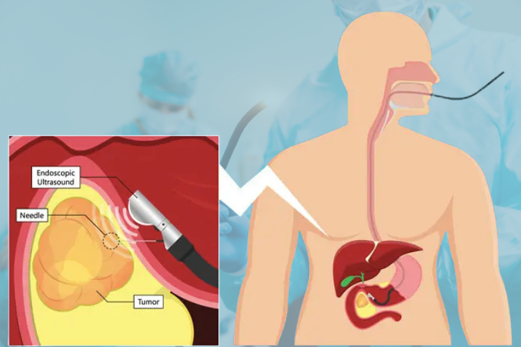
During the procedure, the patient is usually given mild sedation for comfort. The gastroenterologist gently inserts the endoscope either through the mouth (for upper GI EUS) or the rectum (for lower GI EUS). The ultrasound probe then emits high-frequency sound waves that create real-time images on a monitor.
2
EUS not only visualizes the layers of the GI wall but also detects abnormalities in adjacent organs. In many cases, it enables fine-needle aspiration (FNA) or biopsy, where a thin needle is passed through the endoscope to collect tissue samples for laboratory analysis. This minimally invasive approach helps in accurate diagnosis without the need for open surgery.
What to Expect During and After the Procedure
EUS is generally safe and well-tolerated. The procedure typically lasts 30 to 90 minutes depending on whether a biopsy is needed. Patients may experience mild throat discomfort (if upper EUS) or bloating afterward, but these effects usually resolve quickly. Most individuals can resume normal activities within a day.
Potential risks are low but may include minor bleeding at the biopsy site, infection, or rare complications like pancreatitis. Doctors discuss these with patients beforehand to ensure informed consent.
Conclusion
Endoscopic Ultrasound (EUS) represents a powerful tool in modern gastroenterology and surgical gastroenterology. By merging endoscopic visualization with high-precision ultrasound, it delivers detailed insights into the digestive system and surrounding structures, enabling timely and accurate diagnosis of a wide range of conditions. For patients dealing with complex GI issues, pancreatic disorders, or suspected malignancies, EUS offers a minimally invasive path to better understanding and managing their health. Consulting an experienced specialist in advanced endoscopic procedures ensures optimal outcomes and personalized care.